Что такое findslide.org?

FindSlide.org - это сайт презентаций, докладов, шаблонов в формате PowerPoint.


Для правообладателей

Обратная связь

Email: Нажмите что бы посмотреть 

Яндекс.Метрика

Презентация на тему Introduction. Essential Cytology

Содержание

Histology studies the organization of the tissues and organs of the body.Cytology studies the structure and functions of the cell. Embryology researches embryonic development (formation) of the body
Department of Histology, Cytology and EmbryologyLecture 1. Introduction. Histology studies the organization of the tissues and organs of the body.Cytology Cytology Note:1. The cell is the smallest structural and functional unit of the Types of cells in human body Cells produce matrix Methods of investigation Microscopy – basic methodLight microscope: Histological slide: Electron microscopy Electron microscopy researches Ultrastructure of cells (organelles) and organisation of intercellular matrix Light and electron microscopy - are 2 mane methods in histology Levels of biological systemsBiomolecules Membranes Phospholipids structure :Phosphate group (hydrophilic heads)GlycerolFatty acids (hydrophobic tails) Membrane contents:A. Phospholipids: (1 – hydrophilic head, 2 – hydrophobic tails)B. (3 Lipids   may be:Phospholipids – triglycerides (polar)Cholesterol (non-polar) Proteins   may constitute close to 50% of membrane content Proteins  function:  1- channels, 2- pumps,3- receptors, 4- enzymes, 5- integrative, 6- structural Carbohydrates Present in the outer cell membraneForm Receptors Outer cell membrane – cytolemma or plasmalemma Membranes form:Outer cell membraneOrganellesVesicles Nuclear envelop Cell consists of: - Outer cell membrane, - Cytoplasm and - Nucleus 12GIf cells contact, outer cell membrane forms junctions Types of Cell junctionTight  junctionGap junctionDesmosomes Tight junction prevents the movement of molecules into the intercellular spaces present between epithelial cells Gap junctionchannels between cells Desmosomes -Provide cell attachment Inside the cell …Cytoplasm consists of:Matrix (hialoplasm, cytozol)OrganellesInclusions Inclusions - granules with secretions, pigment granules, lipid and glycogen droplets Organelles: (classification by structure)Membranous Non-membranous Organelles:  (classification by function)General (present in every cell, perform general function)Ex.: Rough endoplasmic reticulumMembranes form a network of sac-like structures called cisternae .Ribosomes Smooth endoplasmic reticulum, SER SER structure: membranes form tubules without ribosomes. Function: 1. synthesizis of lipids.2. Golgi complex (or apparatus)= a pack of sacs. Golgi complex …… is connected with endoplasmic reticulum Golgi apparatus  functions: 1. formation of compound molecules – glycoproteins, lipoproteins. Mitochondrion Structure : Contains outer and inner membranes --Folds of inner membrane Mitochondrion Produce ATP molecules (energy) by Krebs cycle LysosomeLysosomes are round vesicles that contain enzymesThese enzymes break down waste materials Non-membranous organelles: MicrofilamentsMicrotubules Centrioles (Cell Center)Ribosomes Note: Microfilaments, Microtubules  form “Skeleton” of the cell Cell centerConsists of 2 centriolesCentriole = 9 x 3 = 27 microtubules;Function Nucleus  consists of:Nucleolemma = nuclear envelope NucleoplasmNucleolusChromatin Nuclear envelope - Consists of two membranes: outer and inner In the nuclear envelope there are gaps, called nuclear pores, providetransport from nucleus into cytoplasm Nuclear pore structure NucleolusNucleolus is the site of active synthesis of ribosomal RNA and formation of ribosomes. Chromatin is the combination of DNA and proteins that make up the Chromatin =  DNA in non-dividing cells. 2 types: 1. heterochromatin (non-active) Euchromatin predominates in metabolically active nuclei, Heterochromatin predominates in metabolically inactive nuclei Chromosome  - is an organized structure of DNA and protein found in dividing cells. Cell cycle The life of a somatic cell is a cyclic process It is Interphase Interphase is a period between two divisions of the cell. Consists In G1 phase: cell grows, performs its routine functions. S- phase   (S- synthesis) DNA molecules are duplicated NOTE: At G2 phaseIn this phase synthesis of proteins, which are required for cell G0 phase cell can leave the cycle and enter to so-called G0 Mitosis  is the process of somatic cells division. Mitosis consists of Prophase Chromosomes become recognisable.the nuclear membrane breaks down and the nucleoli disappear Two centrioles separate and move to opposite poles of the cell. microtubules Metaphase- chromosomes move to a position midway between the two centrioles (the Anaphase - the chromatids separate and move to opposite poles of the Telophase two daughter nuclei are formed chromosomes become indistinct. Nucleoli reappear. Another methods:Polarized microscopy (property of tissues: can rotate the angle of the Gap junctionConsists of six connexin proteins, interacting to form a cylinder with
Слайды презентации

Слайд 2
Histology studies the organization of the tissues and

Histology studies the organization of the tissues and organs of the

organs of the body.

Cytology studies the structure and functions

of the cell.

Embryology researches embryonic development (formation) of the body


Слайд 3 Cytology


Cytology

Слайд 4 Note:
1. The cell is the smallest structural and

Note:1. The cell is the smallest structural and functional unit of

functional unit of the body

2. Cells form

tissues.


3. Tissues form organs and systems
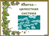

Слайд 5 Types of cells in human body

Types of cells in human body

Слайд 6 Cells produce matrix

Cells produce matrix

Слайд 7 Methods of investigation

Methods of investigation

Слайд 8 Microscopy – basic method
Light microscope:


Histological slide:

Microscopy – basic methodLight microscope: Histological slide:

Слайд 9 Electron microscopy

Electron microscopy

Слайд 10 Electron microscopy researches
Ultrastructure of cells (organelles) and

Electron microscopy researches Ultrastructure of cells (organelles) and organisation of intercellular matrix

organisation of intercellular matrix


Слайд 11 Light and electron microscopy - are 2 mane methods

Light and electron microscopy - are 2 mane methods in histology

in histology


Слайд 12 Levels of biological systems

Biomolecules

Membranes

Levels of biological systemsBiomolecules Membranes    Organelles

Organelles

CELL


Слайд 13 Phospholipids structure :
Phosphate group (hydrophilic heads)
Glycerol
Fatty acids (hydrophobic

Phospholipids structure :Phosphate group (hydrophilic heads)GlycerolFatty acids (hydrophobic tails)

tails)


Слайд 14 Membrane contents:
A. Phospholipids: (1 – hydrophilic head, 2

Membrane contents:A. Phospholipids: (1 – hydrophilic head, 2 – hydrophobic tails)B.

– hydrophobic tails)
B. (3 ) – proteins
C. (4 )

– carbohydrates (only outer cell membrane)

Слайд 15 Lipids may be:
Phospholipids – triglycerides (polar)
Cholesterol (non-polar)

Lipids  may be:Phospholipids – triglycerides (polar)Cholesterol (non-polar)

Слайд 16 Proteins may constitute close to 50% of membrane

Proteins  may constitute close to 50% of membrane content

content


Слайд 17 Proteins function:
1- channels,
2- pumps,
3- receptors,
4-

Proteins function: 1- channels, 2- pumps,3- receptors, 4- enzymes, 5- integrative, 6- structural

enzymes,
5- integrative,
6- structural


Слайд 18 Carbohydrates
Present in the outer cell membrane

Form Receptors

Carbohydrates Present in the outer cell membraneForm Receptors

Слайд 19

Outer cell membrane – cytolemma or plasmalemma

Outer cell membrane – cytolemma or plasmalemma

Слайд 20 Membranes form:
Outer cell membrane
Organelles
Vesicles
Nuclear envelop

Membranes form:Outer cell membraneOrganellesVesicles Nuclear envelop

Слайд 21 Cell consists of:
- Outer cell membrane,
-

Cell consists of: - Outer cell membrane, - Cytoplasm and - Nucleus

Cytoplasm and
- Nucleus


Слайд 22 1
2
G


If cells contact, outer cell membrane forms junctions

12GIf cells contact, outer cell membrane forms junctions

Слайд 23 Types of Cell junction
Tight junction
Gap junction
Desmosomes

Types of Cell junctionTight junctionGap junctionDesmosomes

Слайд 24 Tight junction
prevents the movement of molecules into

Tight junction prevents the movement of molecules into the intercellular spaces present between epithelial cells

the intercellular spaces
present between epithelial cells


Слайд 26 Gap junction
channels between cells


Gap junctionchannels between cells

Слайд 27 Desmosomes -
Provide cell attachment

Desmosomes -Provide cell attachment

Слайд 28 Inside the cell …

Cytoplasm consists of:
Matrix (hialoplasm, cytozol)
Organelles
Inclusions

Inside the cell …Cytoplasm consists of:Matrix (hialoplasm, cytozol)OrganellesInclusions

Слайд 29 Inclusions -
granules with secretions, pigment granules, lipid

Inclusions - granules with secretions, pigment granules, lipid and glycogen droplets

and glycogen droplets


Слайд 30 Organelles: (classification by structure)
Membranous

Non-membranous

Organelles: (classification by structure)Membranous Non-membranous

Слайд 31 Organelles: (classification by function)
General
(present in every cell,

Organelles: (classification by function)General (present in every cell, perform general function)Ex.:

perform general function)

Ex.: Mitochondrion
Special
(in specialised cell, perform special

function)

= Myofibril

Слайд 32 Rough endoplasmic reticulum
Membranes form a network of sac-like

Rough endoplasmic reticulumMembranes form a network of sac-like structures called cisternae

structures called cisternae .
Ribosomes lie on the outer surface.
Function

- synthesis of proteins


Слайд 34 Smooth endoplasmic reticulum, SER

Smooth endoplasmic reticulum, SER

Слайд 35
SER structure: membranes form tubules without ribosomes.
Function:

SER structure: membranes form tubules without ribosomes. Function: 1. synthesizis of


1. synthesizis of lipids.
2. metabolism of carbohydrates

3. drug detoxification

(in liver cells).

4 storage of Ca-ions (only in muscle cell)

Слайд 36 Golgi complex (or apparatus)


= a pack of sacs.

Golgi complex (or apparatus)= a pack of sacs.

Слайд 37 Golgi complex …
… is connected with endoplasmic reticulum

Golgi complex …… is connected with endoplasmic reticulum


Слайд 38 Golgi apparatus functions:
1. formation of compound molecules

Golgi apparatus functions: 1. formation of compound molecules – glycoproteins, lipoproteins.

– glycoproteins, lipoproteins.
2. production of lysosomes and secretory

vesicles.


Слайд 39 Mitochondrion

Structure :
Contains outer and inner membranes

Mitochondrion Structure : Contains outer and inner membranes --Folds of inner


--Folds of inner membrane – cristae
--- Inside

lie matryx

Слайд 40 Mitochondrion

Produce ATP molecules (energy) by Krebs cycle

Mitochondrion Produce ATP molecules (energy) by Krebs cycle


Слайд 41 Lysosome
Lysosomes are round vesicles that contain enzymes
These enzymes

LysosomeLysosomes are round vesicles that contain enzymesThese enzymes break down waste

break down waste materials and cellular debris and digest

the materials within phagosomes.


Слайд 42 Non-membranous organelles:
Microfilaments
Microtubules
Centrioles (Cell Center)

Ribosomes

Non-membranous organelles: MicrofilamentsMicrotubules Centrioles (Cell Center)Ribosomes

Слайд 43 Note:
Microfilaments, Microtubules form “Skeleton” of the cell

Note: Microfilaments, Microtubules form “Skeleton” of the cell

Слайд 44 Cell center
Consists of 2 centrioles

Centriole = 9 x

Cell centerConsists of 2 centriolesCentriole = 9 x 3 = 27

3 = 27 microtubules;

Function - formation of mitotic spindle


Слайд 45 Nucleus consists of:
Nucleolemma = nuclear envelope
Nucleoplasm

Nucleolus
Chromatin

Nucleus consists of:Nucleolemma = nuclear envelope NucleoplasmNucleolusChromatin

Слайд 46 Nuclear envelope
- Consists of two membranes:
outer

Nuclear envelope - Consists of two membranes: outer and inner

and inner


Слайд 47 In the nuclear envelope
there are gaps, called

In the nuclear envelope there are gaps, called nuclear pores, providetransport from nucleus into cytoplasm

nuclear pores, provide
transport from nucleus into cytoplasm


Слайд 48 Nuclear pore structure

Nuclear pore structure

Слайд 49 Nucleolus
Nucleolus is the site of active synthesis of

NucleolusNucleolus is the site of active synthesis of ribosomal RNA and formation of ribosomes.

ribosomal RNA and formation of ribosomes.


Слайд 50 Chromatin
is the combination of DNA and proteins

Chromatin is the combination of DNA and proteins that make up

that make up the contents of the nucleus of

a cell.

Слайд 51 Chromatin = DNA in non-dividing cells.

2 types:

Chromatin = DNA in non-dividing cells. 2 types: 1. heterochromatin (non-active)

1. heterochromatin (non-active) - very tightly packed fibrils .


2.euchromatin - active – less condensed chromatin fibrils loops


Слайд 52
Euchromatin predominates in metabolically active nuclei,

Heterochromatin

Euchromatin predominates in metabolically active nuclei, Heterochromatin predominates in metabolically inactive nuclei

predominates in metabolically inactive nuclei


Слайд 53 Chromosome - is an organized structure of DNA

Chromosome - is an organized structure of DNA and protein found in dividing cells.

and protein found in dividing cells.


Слайд 54 Cell cycle

Cell cycle

Слайд 55 The life of a somatic cell is a

The life of a somatic cell is a cyclic process It

cyclic process
It is called cell cycle
It consists

of two periods: interphase and mitosis.

Слайд 56 Interphase
Interphase is a period between two divisions

Interphase Interphase is a period between two divisions of the cell.

of the cell.
Consists of 3 phases - G1

, S , G2


Слайд 57 In G1 phase:
cell grows, performs its routine

In G1 phase: cell grows, performs its routine functions.

functions.


Слайд 58 S- phase (S- synthesis)
DNA molecules are

S- phase  (S- synthesis) DNA molecules are duplicated NOTE: At

duplicated

NOTE: At the beginning of this phase the

chromosome number is 2N
and at the end each chromosome consists of two DNA molecules or two chromatids, the chromosome number is 4N.


Слайд 59 G2 phase
In this phase synthesis of proteins, which

G2 phaseIn this phase synthesis of proteins, which are required for

are required for cell division, takes place.

After phase

G2 mitosis always begins

Слайд 60 G0 phase
cell can leave the cycle and

G0 phase cell can leave the cycle and enter to so-called

enter to so-called G0 phase (outside the cycle). They

are reserve or stem cell.


Слайд 61 Mitosis is the process of somatic cells division.

Mitosis

Mitosis is the process of somatic cells division. Mitosis consists of

consists of four phase:
prophase,
metaphase,
anaphase,
telophase.


Слайд 62 Prophase
Chromosomes become recognisable.
the nuclear membrane breaks down

Prophase Chromosomes become recognisable.the nuclear membrane breaks down and the nucleoli disappear

and the nucleoli disappear


Слайд 63 Two centrioles separate and move to opposite poles

Two centrioles separate and move to opposite poles of the cell.

of the cell.

microtubules pass from one centriole to

other and form a spindle of division.


Слайд 64 Metaphase
- chromosomes move to a position midway between

Metaphase- chromosomes move to a position midway between the two centrioles

the two centrioles (the equator of the cell) and

form the equatorial plate


Слайд 65 Anaphase
- the chromatids separate and move to

Anaphase - the chromatids separate and move to opposite poles of

opposite poles of the cell
At the end of anaphase

chromatids are called chromosomes.


Слайд 66 Telophase
two daughter nuclei are formed
chromosomes become

Telophase two daughter nuclei are formed chromosomes become indistinct. Nucleoli reappear.

indistinct.
Nucleoli reappear.


Слайд 67 Another methods:
Polarized microscopy (property of tissues: can rotate

Another methods:Polarized microscopy (property of tissues: can rotate the angle of

the angle of the plan of polarized light)

Faso-contrast microscopy


  • Имя файла: introduction-essential-cytology.pptx
  • Количество просмотров: 109
  • Количество скачиваний: 0