- Главная
- Разное
- Бизнес и предпринимательство
- Образование
- Развлечения
- Государство
- Спорт
- Графика
- Культурология
- Еда и кулинария
- Лингвистика
- Религиоведение
- Черчение
- Физкультура
- ИЗО
- Психология
- Социология
- Английский язык
- Астрономия
- Алгебра
- Биология
- География
- Геометрия
- Детские презентации
- Информатика
- История
- Литература
- Маркетинг
- Математика
- Медицина
- Менеджмент
- Музыка
- МХК
- Немецкий язык
- ОБЖ
- Обществознание
- Окружающий мир
- Педагогика
- Русский язык
- Технология
- Физика
- Философия
- Химия
- Шаблоны, картинки для презентаций
- Экология
- Экономика
- Юриспруденция
Что такое findslide.org?
FindSlide.org - это сайт презентаций, докладов, шаблонов в формате PowerPoint.
Обратная связь
Email: Нажмите что бы посмотреть
Презентация на тему Optical identification using imperfections in 2D materials
Содержание
- 2. IntroductionThe ability to uniquely identify an object
- 3.
- 4. ObjectiveTo analise a proposed simple optical technique to read unique information from nanometer-scale defects in 2D materials.
- 5. TasksMethodResults for WS2 from mechanically exfoliationResults for WS2 from chemical vapor depositionConclusion
- 6. MethodMeasurement apparatus, in which the photoluminescence from
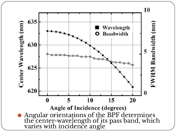
- 7. Angular orientations of the BPF determines the
- 8. Concept of the angular selective transmissionChanging the
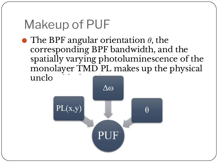
- 9. Makeup of PUFThe BPF angular orientation θ, the
- 10. Results for WS2 from mechanically exfoliation
- 11. Results for WS2 from chemical vapor depositionAngular-dependent
- 12. Angular dependent PL images of WS2 monolayer
- 13. Скачать презентацию
- 14. Похожие презентации
IntroductionThe ability to uniquely identify an object or device is important for authentication. Imperfections, locked into structures during fabrication, can be used to provide a fingerprint that is challenging to reproduce.
