- Главная
- Разное
- Бизнес и предпринимательство
- Образование
- Развлечения
- Государство
- Спорт
- Графика
- Культурология
- Еда и кулинария
- Лингвистика
- Религиоведение
- Черчение
- Физкультура
- ИЗО
- Психология
- Социология
- Английский язык
- Астрономия
- Алгебра
- Биология
- География
- Геометрия
- Детские презентации
- Информатика
- История
- Литература
- Маркетинг
- Математика
- Медицина
- Менеджмент
- Музыка
- МХК
- Немецкий язык
- ОБЖ
- Обществознание
- Окружающий мир
- Педагогика
- Русский язык
- Технология
- Физика
- Философия
- Химия
- Шаблоны, картинки для презентаций
- Экология
- Экономика
- Юриспруденция
Что такое findslide.org?
FindSlide.org - это сайт презентаций, докладов, шаблонов в формате PowerPoint.
Обратная связь
Email: Нажмите что бы посмотреть
Презентация на тему Clinical anatomy of the upper limb
Содержание
- 3. Arteries of the upper limb
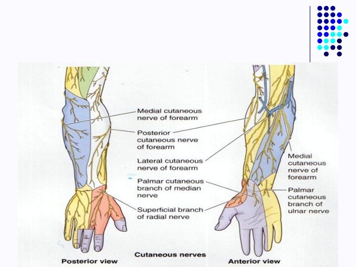
- 4. Skin innervations
- 5. Upper limb
- 6. Regio scapularis
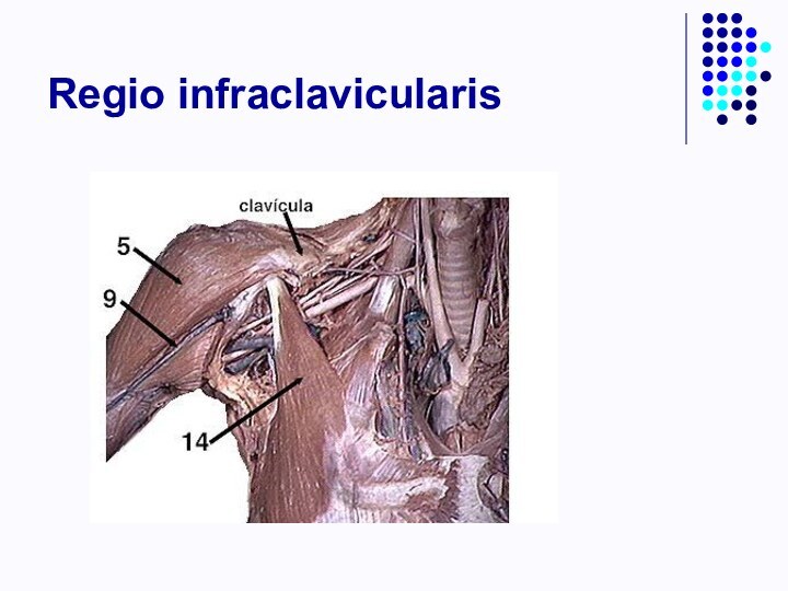
- 9. Regio infraclavicularis
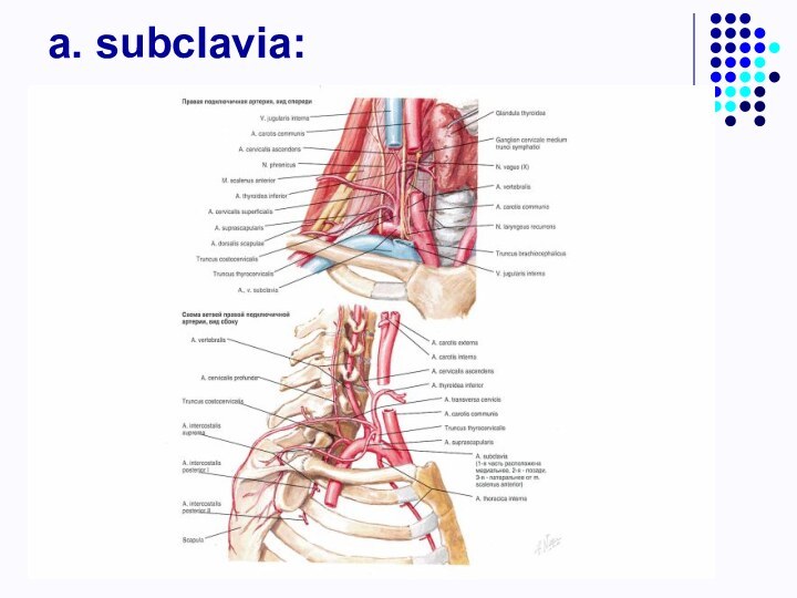
- 12. a. subclavia:
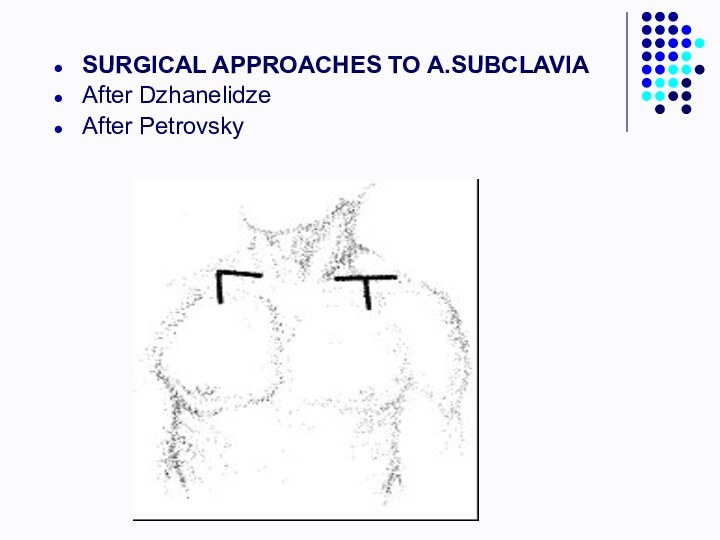
- 14. SURGICAL APPROACHES TO A.SUBCLAVIAAfter DzhanelidzeAfter Petrovsky
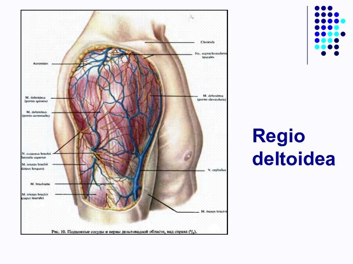
- 15. Regio deltoidea
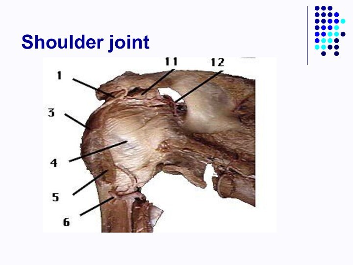
- 16. Shoulder joint
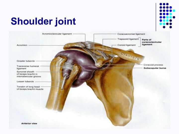
- 17. Shoulder joint
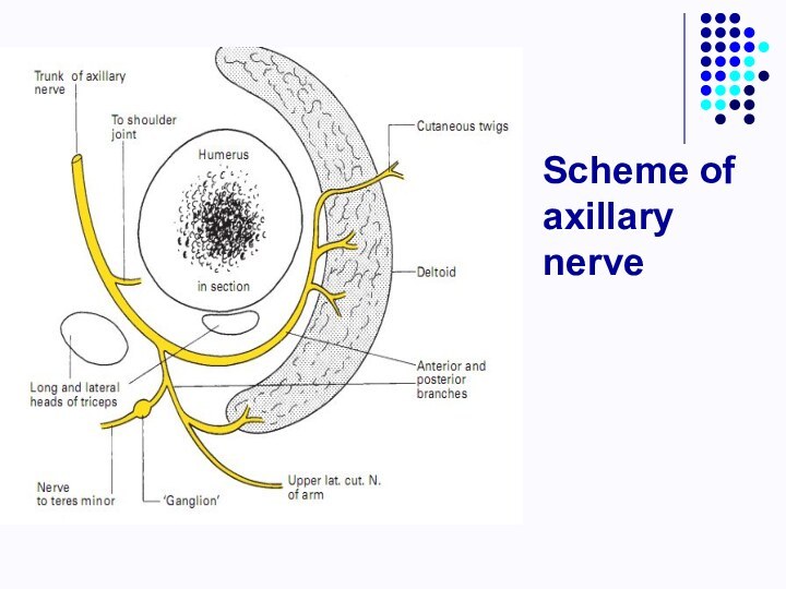
- 18. Scheme of axillary nerve
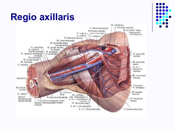
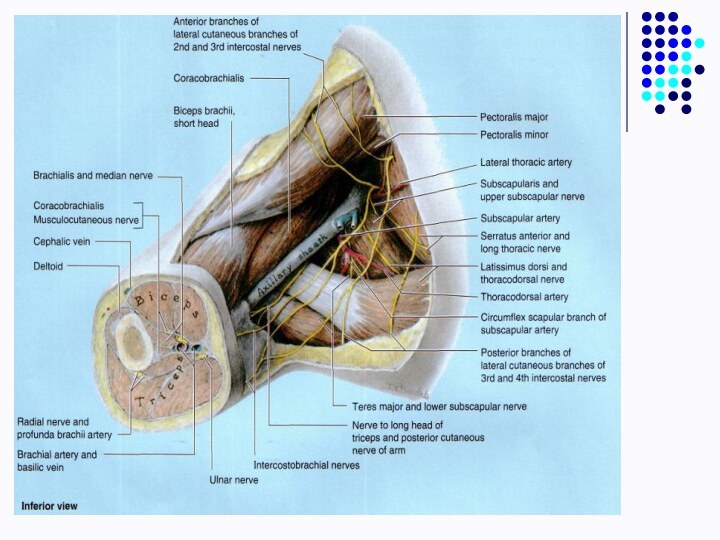
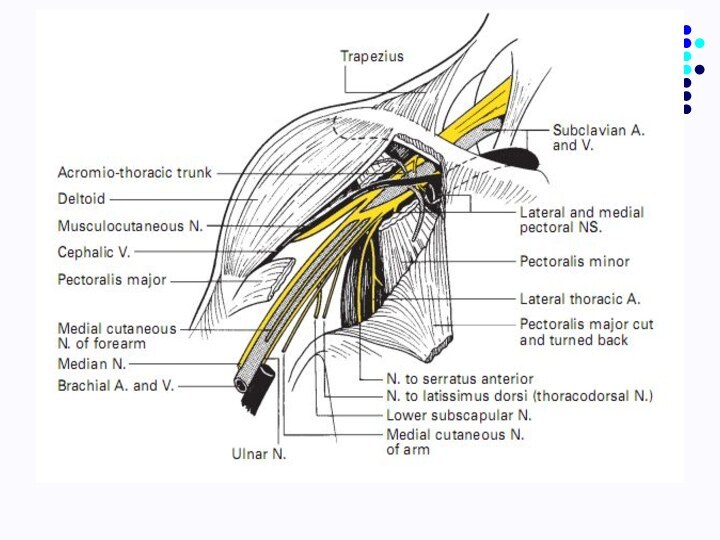
- 19. Regio axillaris
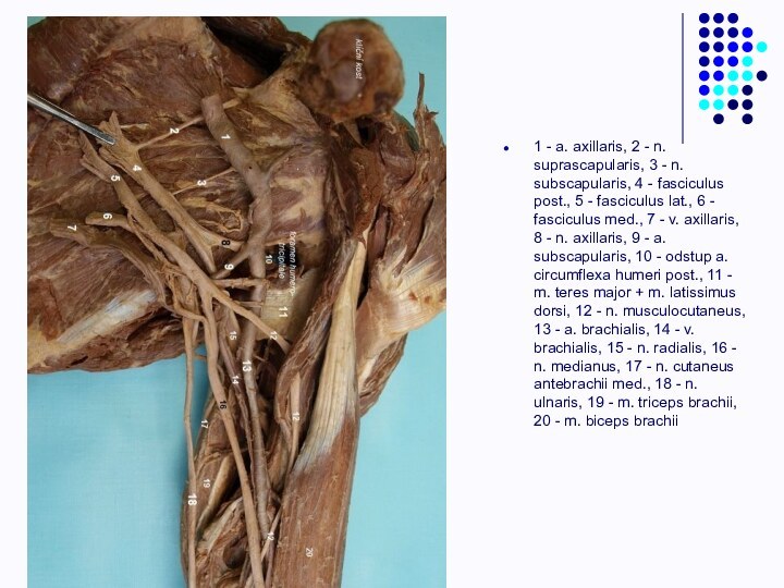
- 23. 1 - a. axillaris, 2 - n.
- 24. Regio brachii posterior
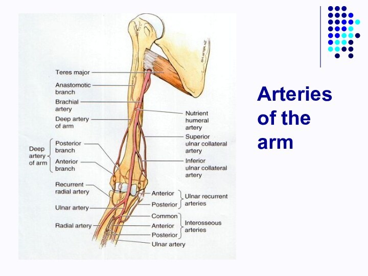
- 25. Arteries of the arm
- 28. Regio cubiti anterior
- 29. Regio cubiti1 - m. biceps brachii, 2
- 30. Fossa cubiti
- 31. Bursas of the elbow joint
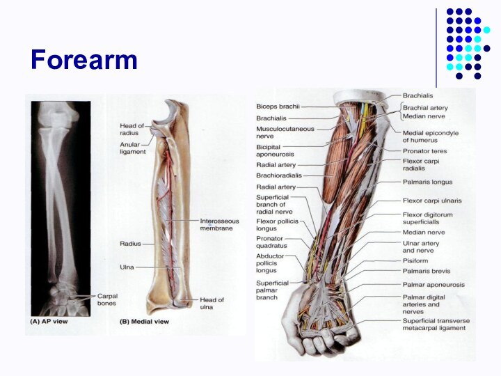
- 32. Forearm
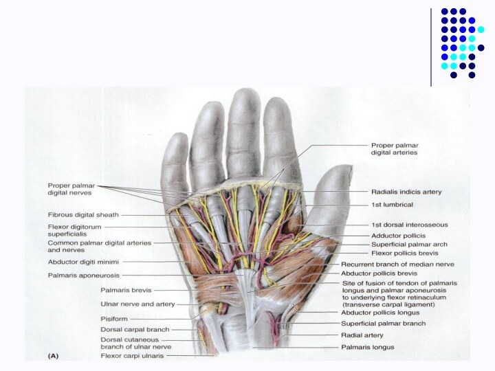
- 33. Topography of the forearm and handAntebrachial grooves:Lateral
- 34. Topography of the forearmCanal of the ulnar
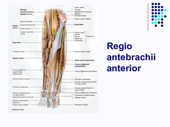
- 35. Regio antebrachii anterior
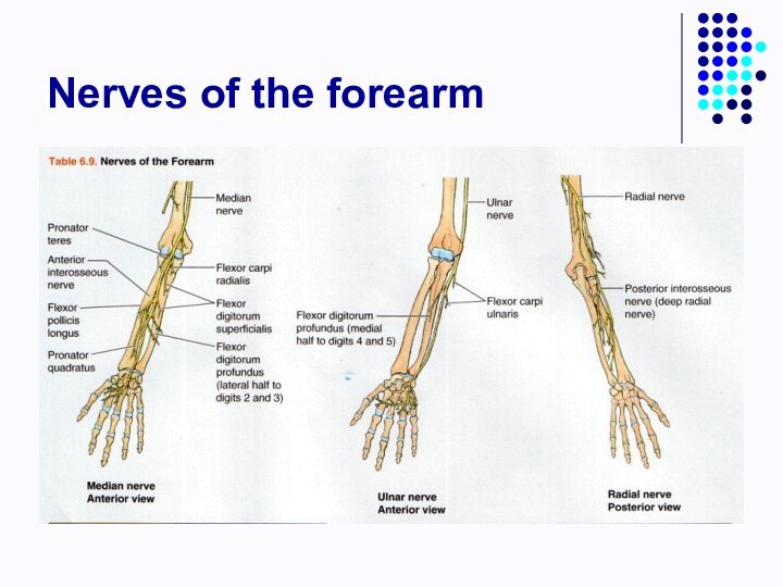
- 37. Nerves of the forearm
- 40. Retinaculi of the upper limbRetinaculum flexorumCanalis carpi
- 41. Retinaculi of the upper limbRetinaculum extensorum/6 canals
- 42. Synovial sheaths of the palmar and dorsal surface of the hand
- 45. Скачать презентацию
- 46. Похожие презентации
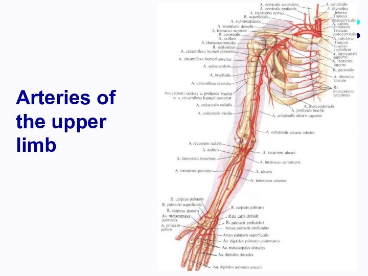
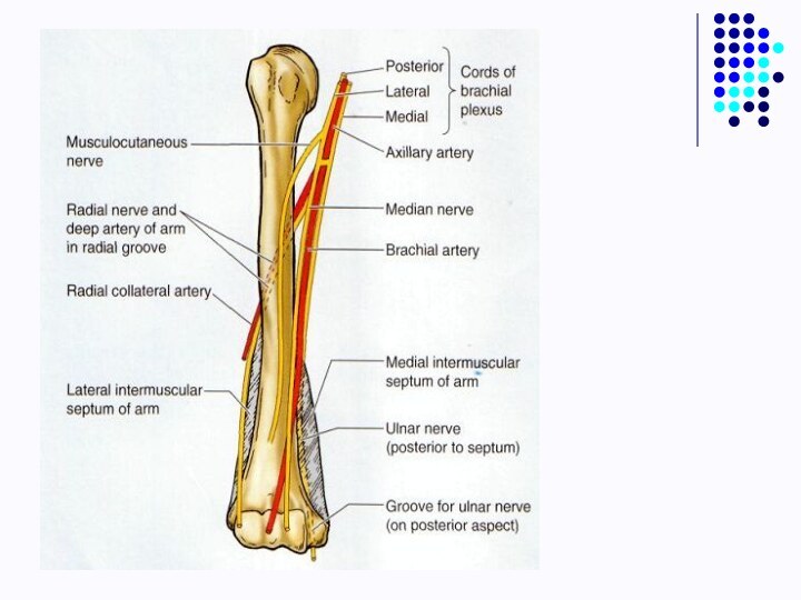
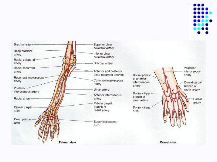
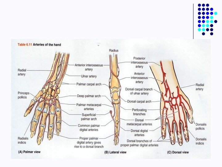
Arteries of the upper limb










Слайд 29
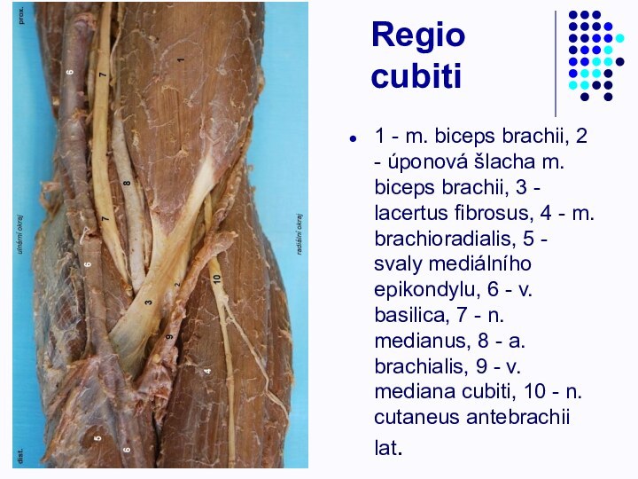
Regio cubiti
1 - m. biceps brachii, 2 -
úponová šlacha m. biceps brachii, 3 - lacertus fibrosus,
4 - m. brachioradialis, 5 - svaly mediálního epikondylu, 6 - v. basilica, 7 - n. medianus, 8 - a. brachialis, 9 - v. mediana cubiti, 10 - n. cutaneus antebrachii lat.
Слайд 33
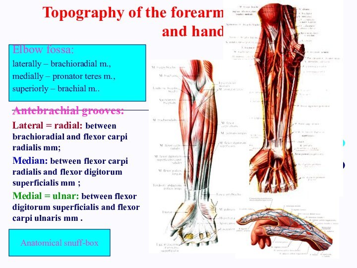
Topography of the forearm
and hand
Antebrachial grooves:
Lateral = radial:
between brachioradial and flexor carpi radialis mm;
Median: between flexor
carpi radialis and flexor digitorum superficialis mm ;Medial = ulnar: between flexor digitorum superficialis and flexor carpi ulnaris mm .
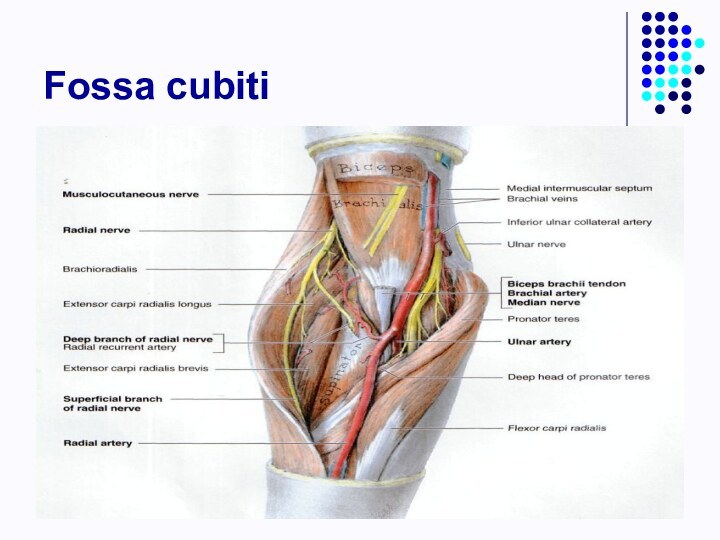
Elbow fossa:
laterally – brachioradial m.,
medially – pronator teres m.,
superiorly – brachial m..
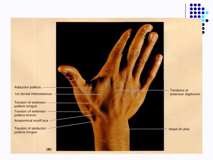
Anatomical snuff-box
Слайд 34
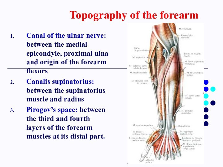
Topography of the forearm
Canal of the ulnar nerve:
between the medial epicondyle, proximal ulna and origin of
the forearm flexorsCanalis supinatorius: between the supinatorius muscle and radius
Pirogov’s space: between the third and fourth layers of the forearm muscles at its distal part.
Слайд 40
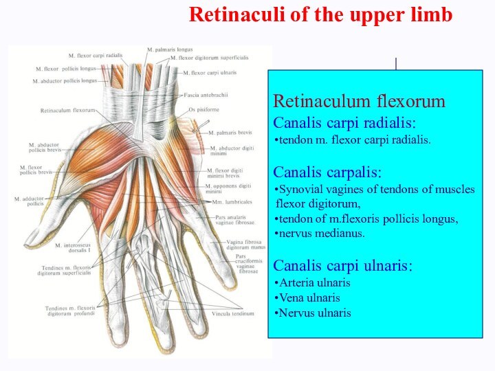
Retinaculi of the upper limb
Retinaculum flexorum
Canalis carpi radialis:
tendon m. flexor carpi radialis.
Canalis carpalis:
Synovial vagines
of tendons of musclesflexor digitorum,
tendon of m.flexoris pollicis longus,
nervus medianus.
Canalis carpi ulnaris:
Arteria ulnaris
Vena ulnaris
Nervus ulnaris
Слайд 41
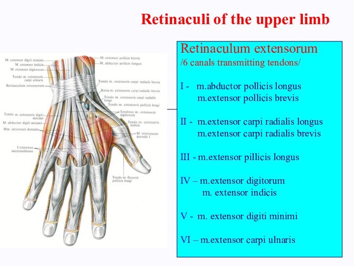
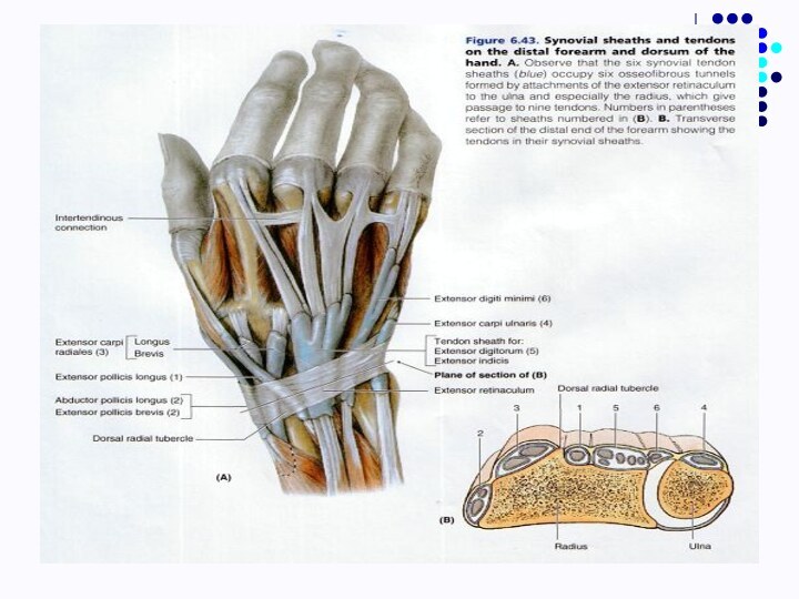
Retinaculi of the upper limb
Retinaculum extensorum
/6 canals transmitting
tendons/
I - m.abductor pollicis longus
m.extensor pollicis brevisII - m.extensor carpi radialis longus
m.extensor carpi radialis brevis
III - m.extensor pillicis longus
IV – m.extensor digitorum
m. extensor indicis
V - m. extensor digiti minimi
VI – m.extensor carpi ulnaris


