- Главная
- Разное
- Бизнес и предпринимательство
- Образование
- Развлечения
- Государство
- Спорт
- Графика
- Культурология
- Еда и кулинария
- Лингвистика
- Религиоведение
- Черчение
- Физкультура
- ИЗО
- Психология
- Социология
- Английский язык
- Астрономия
- Алгебра
- Биология
- География
- Геометрия
- Детские презентации
- Информатика
- История
- Литература
- Маркетинг
- Математика
- Медицина
- Менеджмент
- Музыка
- МХК
- Немецкий язык
- ОБЖ
- Обществознание
- Окружающий мир
- Педагогика
- Русский язык
- Технология
- Физика
- Философия
- Химия
- Шаблоны, картинки для презентаций
- Экология
- Экономика
- Юриспруденция
Что такое findslide.org?
FindSlide.org - это сайт презентаций, докладов, шаблонов в формате PowerPoint.
Обратная связь
Email: Нажмите что бы посмотреть
Презентация на тему Hysterosalpingography (HSG)
Содержание
- 2. Hysterosalpingography (HSG) is a radiologic procedure to
- 3. PROCEDURE The procedure involves X-rays. It should
- 4. A hysterosalpingogram. Note the catheter entering at
- 6. HOW IS HSG DONE? HSG is done
- 7. The procedure is performed as follows:1. You
- 8. What are the risks associated with HSG?Severe
- 16. Скачать презентацию
- 17. Похожие презентации
Hysterosalpingography (HSG) is a radiologic procedure to investigate the shape of the uterine cavity and the shape and patency of the fallopian tubes. It entails the injection of a radio-opaque material into the cervical canal and

Слайд 3
PROCEDURE
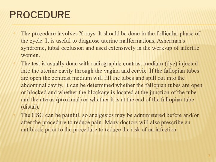
The procedure involves X-rays. It should be done
in the follicular phase of the cycle. It is
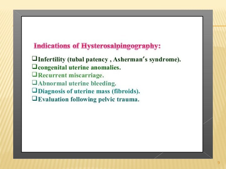
useful to diagnose uterine malformations, Asherman's syndrome, tubal occlusion and used extensively in the work-up of infertile women.The test is usually done with radiographic contrast medium (dye) injected into the uterine cavity through the vagina and cervix. If the fallopian tubes are open the contrast medium will fill the tubes and spill out into the abdominal cavity. It can be determined whether the fallopian tubes are open or blocked and whether the blockage is located at the junction of the tube and the uterus (proximal) or whether it is at the end of the fallopian tube (distal).
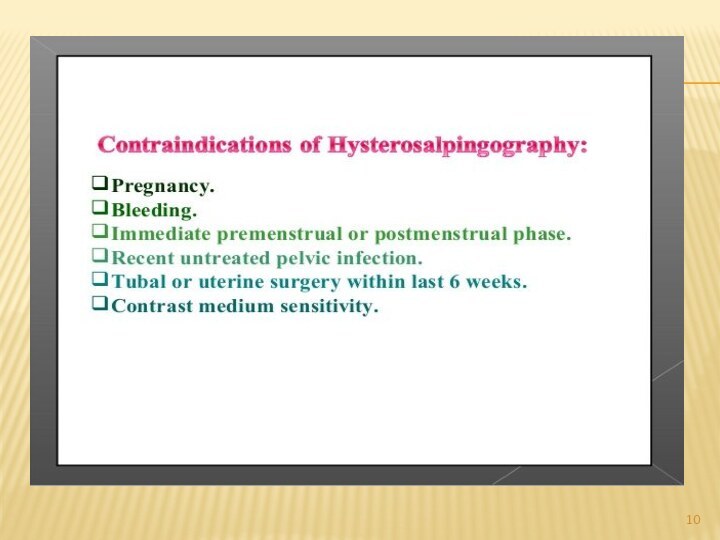
The HSG can be painful, so analgesics may be administered before and/or after the procedure to reduce pain. Many doctors will also prescribe an antibiotic prior to the procedure to reduce the risk of an infection.
Слайд 4
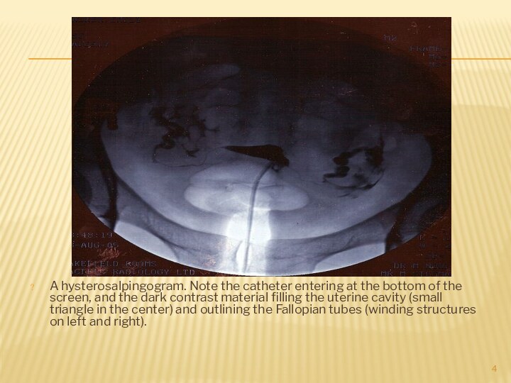
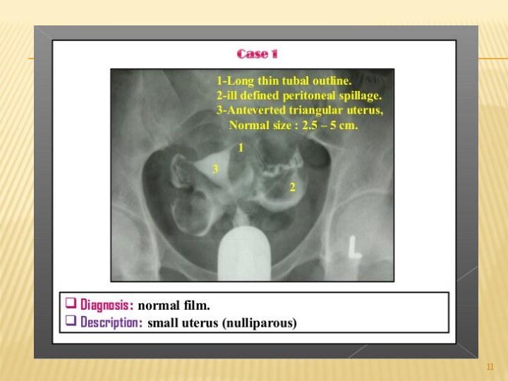
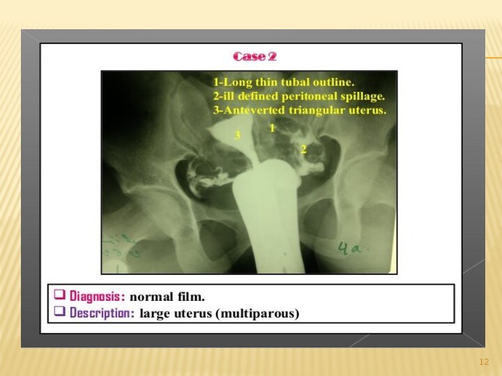
A hysterosalpingogram. Note the catheter entering at the
bottom of the screen, and the dark contrast material
filling the uterine cavity (small triangle in the center) and outlining the Fallopian tubes (winding structures on left and right).
Слайд 6
HOW IS HSG DONE?
HSG is done in a
hospital, clinic, or health care provider’s office. It is
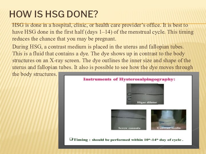
best to have HSG done in the first half (days 1–14) of the menstrual cycle. This timing reduces the chance that you may be pregnant.During HSG, a contrast medium is placed in the uterus and fallopian tubes. This is a fluid that contains a dye. The dye shows up in contrast to the body structures on an X-ray screen. The dye outlines the inner size and shape of the uterus and fallopian tubes. It also is possible to see how the dye moves through the body structures.
Слайд 7
The procedure is performed as follows:
1. You will
be asked to lie on your back with your
feet placed as for a pelvic exam. A device called a speculum is inserted into the vagina. It holds the walls of the vagina apart to allow the cervix to be viewed. The cervix is cleaned.2. The end of the cervix may be injected with local anesthesia (pain relief). You may feel a slight pinch or tug as this is done.
3. One of two methods may be used to insert the dye. In one method, the cervix is grasped with a device to hold it steady.
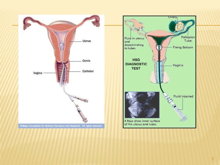
An instrument called a cannula is then inserted into the cervix. In the other method, a thin plastic tube is passed into the cervical opening. The tube has a small balloon at the end that is inflated. The balloon keeps the tube in place in the uterus.
4. The speculum is removed, and you are placed beneath an X-ray machine.
5. The fluid slowly is placed through the cannula or tube into the uterus and fallopian tubes. The fluid may cause cramping.
If the tubes are blocked, the fluid will cause them to stretch.
6. X-ray images are made as the contrast medium fills the uterus and tubes. You may be asked to change position. If there
is no blockage, the fluid will spill slowly out the far ends of the tubes. After it spills out, the fluid is absorbed by the body.
7. After the images are made, the cannula or tube is removed.
Слайд 8
What are the risks associated with HSG?
Severe problems
after an HSG are rare. They include an allergic
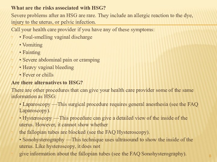
reaction to the dye, injury to the uterus, or pelvic infection.Call your health care provider if you have any of these symptoms:
• Foul-smelling vaginal discharge
• Vomiting
• Fainting
• Severe abdominal pain or cramping
• Heavy vaginal bleeding
• Fever or chills
Are there alternatives to HSG?
There are other procedures that can give your health care provider some of the same information as HSG:
• Laparoscopy —This surgical procedure requires general anesthesia (see the FAQ Laparoscopy).
• Hysteroscopy —This procedure can give a detailed view of the inside of the uterus. However, it cannot show whether
the fallopian tubes are blocked (see the FAQ Hysteroscopy).
• Sonohysterography —This technique uses ultrasound to show the inside of the uterus. Like hysteroscopy, it does not
give information about the fallopian tubes (see the FAQ Sonohysterography).


